Taking Tissue Models to the Final Frontier
By Janelle Weaver
Some may think of outer space as a sterile environment, but infections are a common occurrence aboard spacecraft, where exposure to microgravity negatively affects immune system function. For example, about half of the astronauts who flew in Apollo missions reported minor bacterial or viral infections within a week of their return. Although data for U.S. missions are limited, 26 instances of infection were reported for American astronauts from April 1989 to January 1998.
Space flight is likely to cause deleterious changes to the composition of bacterial flora, leading to an increased risk of infection. The environment may also affect the susceptibility of microorganisms within the spacecraft to antibiotics, key components of flown medical kits, and may modify the virulence of bacteria and other microorganisms that contaminate the fabric of the International Space Station and other flight platforms.
“It has been known since the early days of human space flight that astronauts are more prone to infection,” says Dongeun (Dan) Huh, Wilf Family Term Assistant Professor in Bioengineering at Penn Engineering. “Infections can potentially be a serious threat to astronauts, but we don’t have a good fundamental understanding of how the microgravity environment changes the way our immune system reacts to pathogens.”
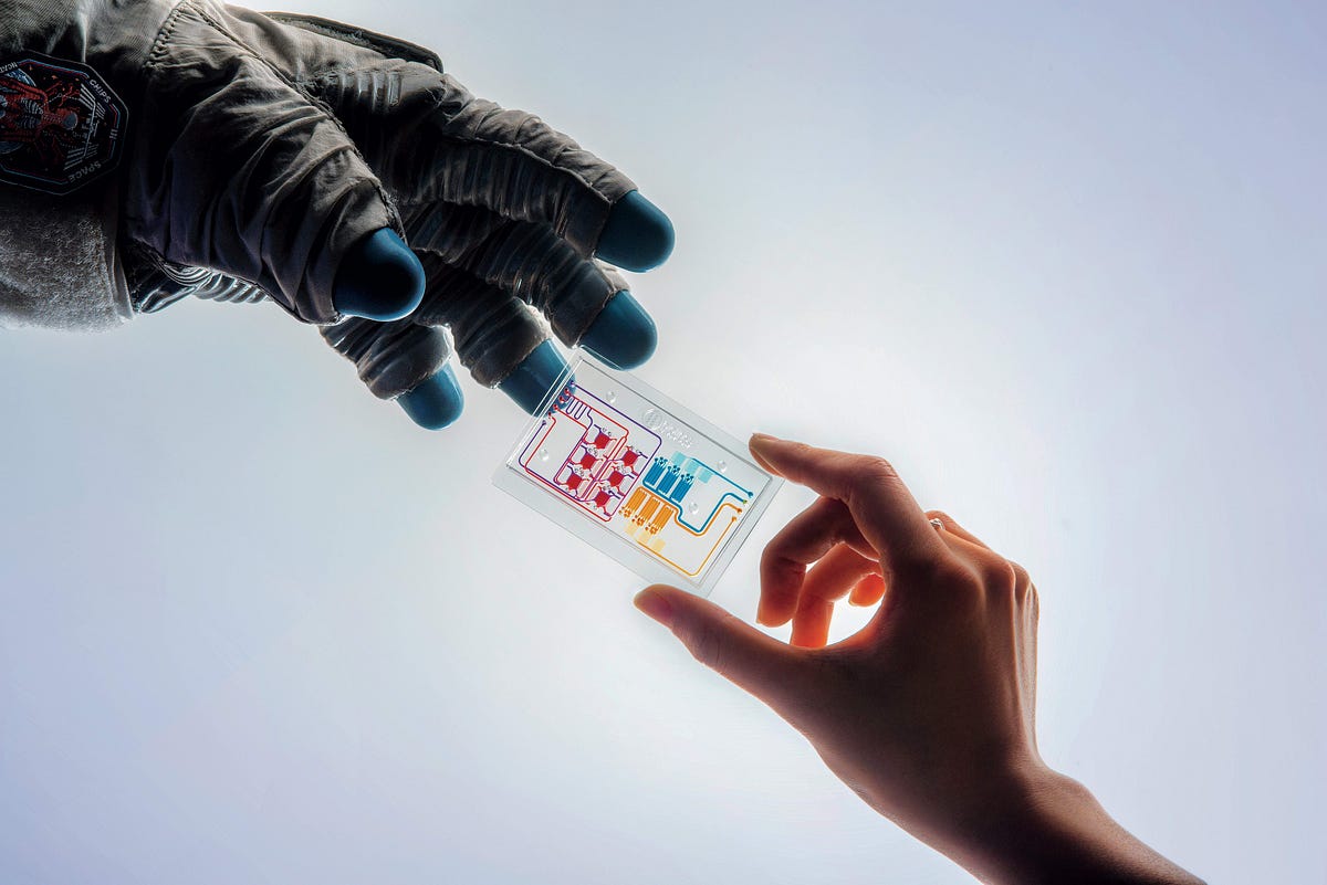
In collaboration with G. Scott Worthen, a physician-scientist in neonatology at the Children’s Hospital of Philadelphia, Huh will attempt to answer this question by sending tissues-on-chips to space. Last June, the Center for the Advancement of Science in Space (CASIS) and the National Center for Advancing Translational Sciences (NCATS), part of the National Institutes of Health (NIH), announced that the duo had received funding to study lung host defense in microgravity at the International Space Station.
Huh and Worthen aim to model respiratory infection, which accounts for more than 30 percent of all infections reported in astronauts. The project’s goals are to test engineered systems that model the airway and bone marrow, a critical organ in the immune system responsible for generating white blood cells, and to combine the models to emulate and understand the integrated immune responses of the human respiratory system in microgravity.
With the initial launch aboard a Cygnus NG-11 rocket scheduled for April 2019, the first phase of the project will focus on simulating infection separately on chips containing lung and bone marrow tissue. If successful, the team will integrate the two devices, which would be launched into space two years later. “Ultimately, if we find abnormalities in how the tissues respond to infection, we might be able to come up with intervention strategies that would inhibit or minimize adverse effects of microgravity,” Huh says.
CAPTURING COMPLEXITY
The idea of creating microengineered models of the human lung arose when Huh was a Wyss Technology Development Fellow at the Wyss Institute for Biologically Inspired Engineering at Harvard University. In 2010, his study led to a prestigious publication in the journal Science. The article described a lung-on-a-chip that mimicked the air-blood interface between an alveolar air sac and small blood vessels called capillaries. The device was integrated with a computer-controlled vacuum to produce cyclic stretching of the tissue-tissue interface to simulate breathing movements.
“We demonstrated for the first time that a microengineered system could mimic complex organ-level functions in the lung such as immune responses to bacteria, toxins and environmental particulates,” Huh says. “This generated a lot of excitement in the field and stimulated a collective effort to develop more physiologically relevant in vitro models using microengineering technologies.”
Standard in vitro systems, which consist of cells grown in static culture media in hard, flat plastic dishes, are rudimentary and do not represent the complexity of the human body. Meanwhile, animal models often do not replicate disease-related processes in humans because of species-specific differences. As a result, approximately 90 percent of drugs that enter clinical trials ultimately fail due to safety or efficacy concerns, costing the pharmaceutical industry more than a decade of research and several billions of dollars per drug candidate.
Unlike traditional systems, 3D organs-on-chips capture the architecture and cellular heterogeneity of tissues found in human organs and mimic the dynamic mechanical and biochemical processes of the surrounding environment. And unlike animal models, they are composed of human cells and are amenable to real-time visual analysis of drug responses. According to unofficial feedback from high-level NIH employees, the Science study changed the way that they thought about in vitro testing. Recognizing the potential of the approach, NCATS launched the Tissue Chip for Drug Screening program in 2012.
Since the seminal study in 2010, Huh and his collaborators have made tremendous progress, developing organ-on-a-chip models for the eyes, gut, placenta, cervix and pancreas to study conditions such as dry eye disease, Crohn’s disease, preterm birth and diabetes. Last June, the Cancer Research Institute announced that Huh received its inaugural Technology Impact Award of $1 million to use microchips to study interactions between cancer and immune cells for the development of immunotherapies.
CHALLENGES ABOVE EARTH
Conducting experiments on Earth is hard enough, but space presents an additional layer of unique challenges. To overcome these hurdles, Huh and Worthen are teaming up with two microgravity research companies called SpacePharma and Space Tango. These implementation partners will be intimately involved in the development of the tissues-on-chips in a space-ready fashion, and in the establishment of remote control and real-time data recovery from the International Space Station.
A major challenge will be to execute several complex experimental steps to infect the cells with microorganisms and monitor their responses for the entire duration of the flight. To address this concern, the team is working closely with their implementation partners to develop a fully automated cell-culture platform with imaging capabilities.
If all goes according to plan, the researchers will analyze cytokines, chemokines and other immune molecules secreted by an airway-on-a-chip infected with Pseudomonas aeruginosa bacteria, which can cause severe illness and even death in humans. The bone-marrow-on-a-chip will be used to simulate the mobilization of immune cells called neutrophils, which are among the first responders that travel from the bone marrow to the site of an infection. Meanwhile, control experiments will take place in a terrestrial facility.
“We can visualize how the lung cells natively slow down and block bacteria by secreting more mucus, and most exciting, we can measure how systemic signaling molecules released by the airway cells recruit white blood cells from the bone marrow by stimulating them to enter the bloodstream and find their way to the infected tissue,” says third-year doctoral student Andrei Georgescu. “By sending these organ-modeling chips to the Space Station while also mirroring the experiments down here on Earth, we’ll be able to closely compare differences and understand changes in the behavior of human cells and pathogenic bacteria during an infection.”
The four other “Chips in Space” research projects funded by CASIS and NCATS will examine human physiology and disease aboard the International Space Station. “This collective effort could make a huge impact on the health of astronauts during and after space flight,” Huh says. “We hope that the lessons we learn will better prepare astronauts to fight sickness as they explore the next frontier.”