‘Calculus III for cells’
Last year, researchers from the University of Pennsylvania revealed surprising insights into how cells respond to surface curvature. Specifically, they investigated how cells respond to cylindrical surfaces, which are common in biology. They found that cells change the static configurations of their shapes and internal structures.
“We think of it as the cells doing calculus; the cells sense and respond to the underlying curvature,” says Kathleen Stebe of Penn’s School of Engineering and Applied Science.
Now, the researchers, led by Stebe and recent engineering graduate Nathan Bade in collaboration with Randall Kamien of the School of Arts and Sciences and Richard Assoian of the Perelman School of Medicine, have published a follow up study that Stebe likens to “calc III” for cells, investigating how cells respond to more complex geometries. The research, which could enable new tools in biology and affect how physicians treat things like vascular disease, has been published in the Biophysical Journal.
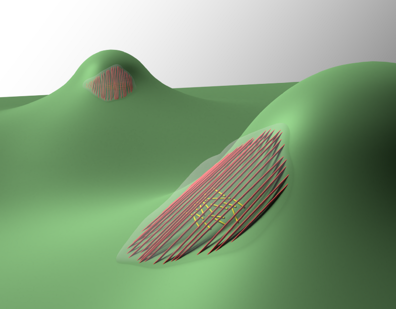
The researchers created a surface other than cylinders that they refer to as a “sphere-with-skirt.” As the name suggests, the upper portion of the surface is spherical, but, as one travels farther down the surface on either side, it forms a skirt that is saddle-like in shape. Because of this, the surface has two non-zero principle curvatures at every point; the spherical portion has what’s called positive Gaussian curvature while the skirt has negative Gaussian curvature.
“We gave this really interesting little mountain to the cells,” Stebe says, “and said, What are you going to do with this smooth mountain that gives you these different curvatures? And it turns out these cells are really clever. They not only change their shapes and internal structures, but they move in dramatically different ways that open new questions about how cells move.”
Cells on stiff surfaces form stress fibers, comprising actin and myosin motors. In the previous study, the researchers found that, surprisingly, on a cylindrical surface the cells actually bend some of the stress fibers along the direction of maximum curvature. Although one population of stress fibers located above the cell’s nucleus aligned along the cylinder axis, another one under the nucleus wrapped around the cylinder circumference. They also found that, by manipulating the ctyoskeleton of the cells, they could recapitulate the alignment pattern of the cytoskeleton that they saw in vivo.
Continue Reading at Penn Today.