Lightweight, tough and strong, our bones are forces of nature. Ottman Tertuliano joined the faculty of Penn Engineering last year to study bone as both a resilient living tissue and sophisticated organic material. AMA Family Assistant Professor in the Department of Mechanical Engineering and Applied Mechanics, Tertuliano creates new tools to measure tissue mechanics at the smallest scales imaginable.
Thanks to his research, we won’t need to imagine. The Tertuliano Lab is hard at work creating visual data that demonstrates how bones behave under dynamic stress — a significant unknown in healthcare.
Whether it be the everyday stress of walking or a more forceful impact, a healthy bone or one compromised by illness, Tertuliano has fine-tuned methods to visualize and explain it. His research has revealed a rich universe of bone’s protective structural mechanisms and his sights are set on thorough characterization and explanation. After an academic year building a lab, training graduate students and running inventive, labor-intensive experiments at Brookhaven National Laboratory, Tertuliano is ready to reap the rewards.
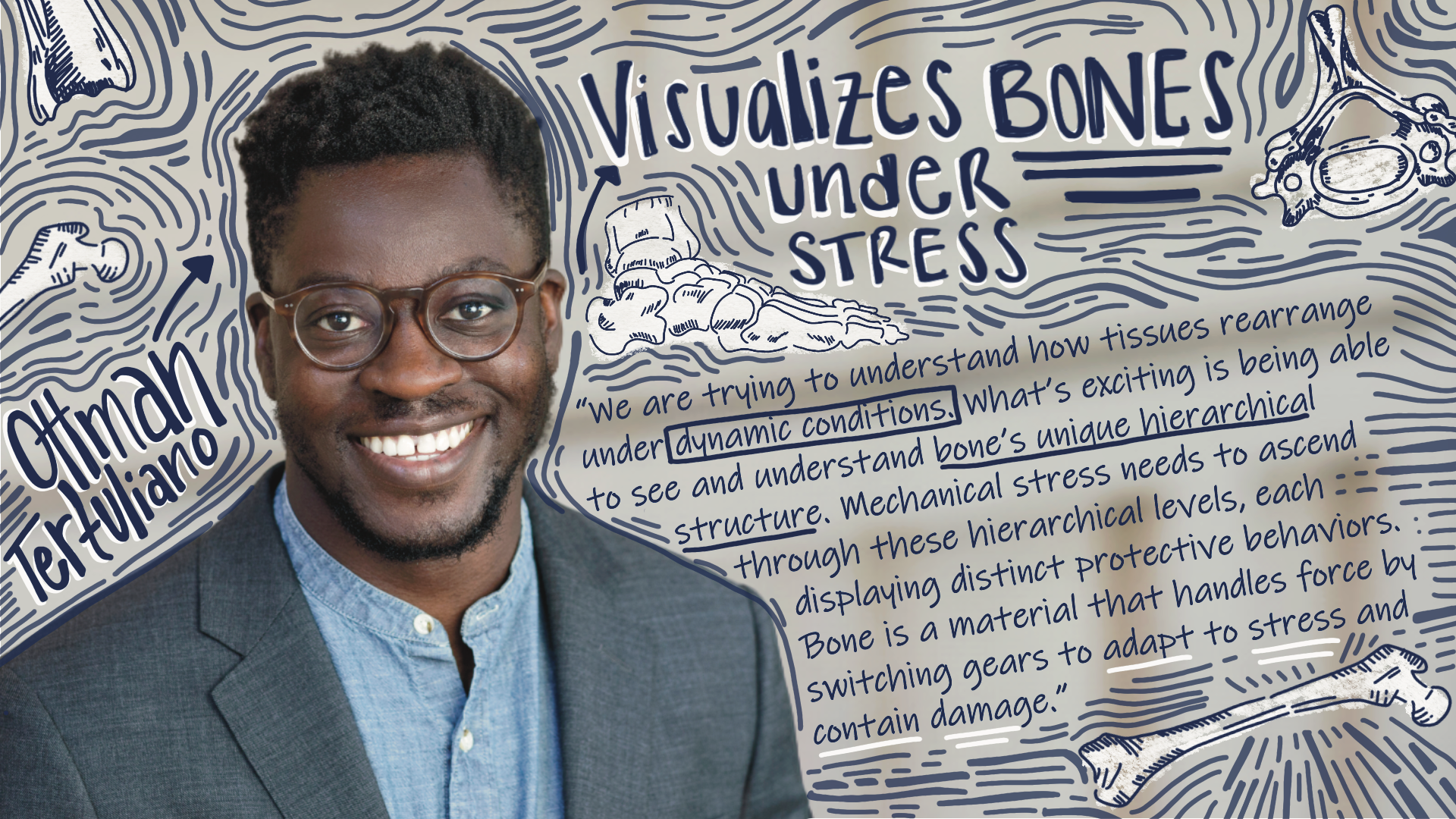
“I’m excited to get to the point where we are collecting really beautiful data. We’re trying to understand how tissues rearrange under dynamic conditions. In just one year — much quicker than I was expecting — we’ve solved a lot of our hardest problems and are ready to dig into some very complicated experiments,” says Tertuliano.
How does bone tissue rearrange? It depends how closely you look. Tertuliano’s research has revealed that bones respond to stress in vastly different ways at different scales.
For example, apply repetitive stress to a bone sample and examine it at the nanoscale. Fracture may occur, but collagen fibers just a few hundred nanometers wide will rearrange themselves to maintain the tissue’s toughness. (For comparison, a human hair measures about 100,000 nanometers wide). These bone-specific, highly mineralized collagen fibers interrupt an expected mechanical failure, fighting to keep the tissue intact. Where a different material might weaken and crumble, bone acts with surprising resilience.
Now examine the sample at a larger length scale — 100 microns or more — and watch a new world of protection appear. Here, a bone’s self-preservation technique looks more like mineralized obstacles deflecting or even arresting a crack as it grows. These defensive barriers built into the very structure of our bones exist in the tissue to overwhelm and stop the fracture, requiring energy to overcome and giving cellular repair the opportunity to begin.
“These are only some of the levels and variety we see in bone behavior,” says Tertuliano. “There are many, many more we are investigating. What’s exciting is being able to see and understand bone’s unique hierarchical structure. Mechanical force needs to work its way through tiered levels, each displaying distinct protective behaviors. Bone is a material that handles force by switching gears, in a sense, to adapt to stress and contain damage.”
Understanding this material will be as valuable to clinicians as to mechanical engineers and materials scientists. The Tertuliano Lab is currently collaborating with radiation oncologists, bioengineers and materials scientists, pursuing insights into the weakening effects of proton radiation cancer treatment on bone tissue and fatigue fracture in metals. The lab’s first two doctoral students, Riti Sharma and Luc Capaldi, anchor the lab’s fruitful diversity, concentrating on bone biomechanics and metals research, respectively.
“The overarching theme of our lab is experimental mechanics. This gives us remarkable tools to study biological systems,” says Sharma, who holds degrees in both Mechanical Engineering and Bioengineering. “On the biological side, we’re working to put human health problems in quantifiable contexts. It’s important to realize that there’s a huge gap between the mechanics of what’s going on in bone and the tools available to clinicians. A clinical X-ray can only tell you so much.”
“The lab is very much bioinspired, focusing on material systems that take their cue from nature,” adds Capaldi, a mechanical engineer working on materials research for aerospace and infrastructure. “Our lab can do mechanical experiments at the nanoscale and microscale, so we can offer small-scale insights into tissues, the mechanics that drive cells to build them, and metal alloys that are inspired by them. From my perspective, natural materials have advantageous properties, and we’d like to be able to understand them better in order to translate these qualities into synthetic form.”
One day, insight into bone’s nanoscale structures may assist orthopedic researchers and practitioners to predict, prevent and contain fracture. These same observations could also help develop materials that are damage-tolerant, super-strong and resistant to failure.
The road forward is paved with experiment, where Tertuliano shines. His passion for experimentation breaks through the everyday grind of research and flourishes under pressure.
“He’s empathetic and supportive when something breaks or doesn’t work, and no one celebrates harder than him when an experiment succeeds,” says Sharma. “He has an extraordinary spirit of persistence and creativity, as well. He’ll try all sorts of unexpected options until something works.”
In the lab’s recent trip to Brookhaven National Laboratory, where they were given a strict 48-hour time window to integrate their customized tool for testing nanoscale bone toughness into an advanced x-ray microscope, Tertuliano took a striking chance.
“Our samples are incredibly delicate, just tiny sections of bone mounted on small wafers. They take a lot of care and effort using tools in the Singh Center’s nanofabrication facilities to prepare. If you turn them wrong, they break, if you breathe on them wrong, they break. But on one of our critical trips to Brookhaven, we discovered our wafers were too large for the X-ray transmission. We had nothing,” says Capaldi.
“Ottman wasn’t fazed. He got out his tools and he cut them. By hand. This is an incredibly small, impossibly fragile object — a one-millimeter-by-one-millimeter wafer with this little 20-micron sample on it — and he just went ahead and willed it to work. He cut five of them and four survived,” says Sharma.
“He cut the samples by hand,” marvels Capaldi. “And we managed to get 3D images of individual collagen fibers in the final 6 hours. It was surreal. It was great science.”