A cavernous, walk-through human heart has entertained 70 years of curiosity, from school trips to international visitors. The Franklin Institute’s Giant Heart is an iconic Philadelphia experience that allows visitors to learn the inner workings of one of our body’s most important muscles from the outside, while weaving through its larger-than-life walls. Such an important Philadelphia teaching tool, the heart was even featured in an episode of Abbott Elementary.
During the Franklin Institute’s 200th Anniversary in 2024, the museum announced that the Giant Heart would temporarily close before re-opening to the public six months later as part of its Body Odyssey exhibit.
Once again, visitors explored complex workings of the human body, discovered the connection between mental and physical health, and experienced the latest advances in medical and sports technology.
The updated exhibit included new ways for visitors to learn about both their bodies, as well as the future of medicine and science, from another iconic Philadelphia institution: The University of Pennsylvania. Several researchers from Penn Medicine’s department of Neurology are featured throughout the exhibit, highlighting futuristic devices and unique career paths.
One iconic Philadelphia institution supporting another
Based on crucial feedback from the Franklin Institute’s test audiences, the new exhibit prioritized looking to the future, in this case, the future of health and technology.
“We wanted to explore where biotechnology is today, where it’s heading in the future, and how it can change our health choices and decisions,” explained Jayatri Das, PhD, Chief Bioscientist and Director of Science Content at the Franklin Institute, and a fellow at Penn’s Center for Neuroscience and Society. “What’s incredible is that these amazing advances in science, medicine, and technology aren’t just happening around the world, they’re happening right here in Philadelphia, and we want to celebrate that.”
In addition to larger-than-life organs and hands-on simulations of robotic surgery and operating a prosthetic hand, the exhibit pays homage to the medical firsts at Penn Medicine that have continued to push the boundaries of science and medicine. From establishing the first hospital in the United States over 270 years ago, to opening the nation’s first medical school over 260 years ago, through more recent breakthroughs like CAR T cell therapy, gene therapy, and the Nobel Prize-winning mRNA vaccine technology, the exhibit illuminates the role of the historic institution in Philadelphia and the world throughout the exhibit with videos, images, and real technology developed by Penn Medicine researchers.
Three of the featured items come from faculty in the Department of Neurology, which was also the first in the nation.
The intersection of biology and engineering
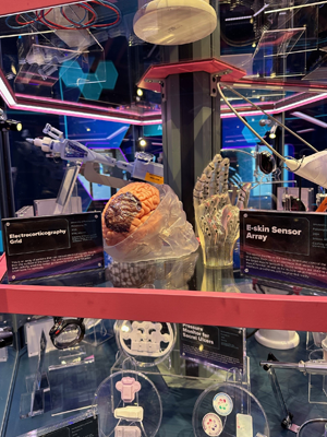
The first is an E-Skin Sensor Array, or a wearable “glove” comprised of dozens of sensors made from a flexible gel-like material, called MXene. The sensors are custom-fabricated for each wearer to fit like a second skin. The device is applied directly to an individual’s skin and can be used both to send signals to the nerves and muscles, as well as receive information on how and when the muscles, nerves, and tendons are engaged.
The sensors are currently used in a study in partnership with Josh Baxter, PhD, an assistant professor of Orthopaedic Surgery, that investigates how individuals recover from Achilles tendinopathy. The sensors are affixed to the calves of people who have injured their Achilles tendon, and send researchers information on how each individual engages their calf muscles and Achilles tendon throughout recovery. Their goal is to understand how individuals move with and without tendon pain, how the muscles surrounding the tendon contribute to pain, and what movements are most impacted. The “glove” sensor array for can be used for similar research in patients with conditions like carpal tunnel syndrome and back pain.
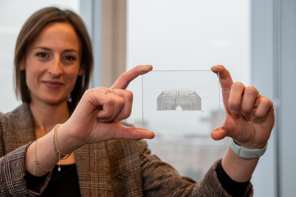
The second sensor Vitale donated to the exhibit is an Electrocorticography Grid, which is a flexible sensor patch that sits on the surface of the brain. While existing brain sensors have one or two electrodes, this patch has 132 electrodes and is flexible enough to follow the ridges of the brain.
Similar to the E-Skin Sensor Arrays, the patch can send electrical stimulation to the brain, like in patients with Parkinson’s disease who receive deep brain stimulation. It can also monitor brain activity, like electroencephalogram (EEG) sensors for patients being monitored for epilepsy. Vitale is currently leading studies to demonstrate the efficacy and safety of this sensor.
“These devices show promise as safe, affordable, and effective tools to listen to the brain tell us how it works, and to intervene when it’s not working properly,” said Vitale. “In the future, we hope to be able to use this sensor to develop all kinds of new and advanced technologies that help us communicate with the brain, from sending targeted impulses that prevent compulsive behavior to transmitting brain signals to control prosthetic devices.”