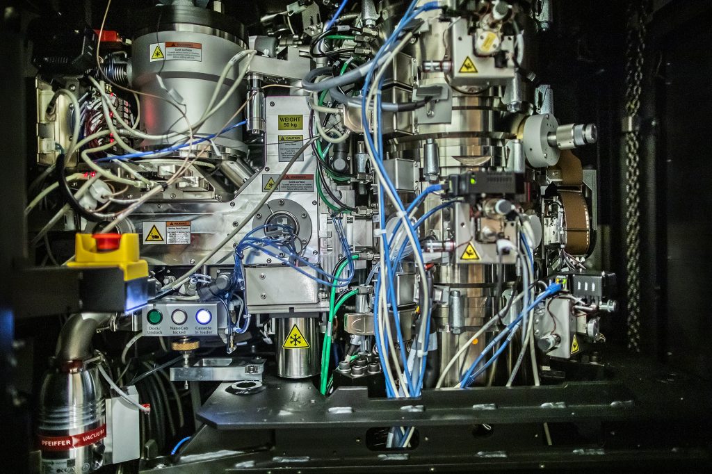
Penn’s Singh Center for Nanotechnology has added the newest cutting-edge technology to its collection of already impressive microscopes: The Krios G3i, a cryogenic electron microscope that will allow researchers to look at cells, proteins, and engineered nanoparticles like never before. The trick is to keep them frozen in time, holding them in their natural environment while bombarding them with ultrafast, ultrahot subatomic particles.
“Cryo-electron microscopy, or cryo-EM, will allow scientists at Penn to understand diseases like cancer, Alzheimer’s, Parkinson’s, and heart and kidney disorders. Biomedical engineers will use cryo-EM to improve nanoparticle technology for drug delivery. Bringing this microscope onto campus is a big deal for everyone,” says Vera Moiseenkova-Bell, associate professor of pharmacology in the Perelman School of Medicine and faculty director of the new Beckman Center for Cryo-Electron Microscopy.
The Arnold and Mabel Beckman Foundation, together with the School of Medicine made this Nobel-winning technology available on Penn’s campus last year, allowing scientists to look at molecules in their natural state at a nearly atomic level.
The ‘cryo revolution’
Moiseenkova-Bell is already seeing firsthand the advantages of using cryo-EM.
“Getting the structures of biologically important molecules at an atomic resolution is now possible because of the cryo revolution. Compared to other techniques, this is so fast. Crystallography can take sometimes over a year to get one structure. Cryo-EM allows you to get a structure in days.”
Many structural biologists like Moiseenkova-Bell consider cryo-EM to be a revolution in their field because of the ways it both supersedes and complements other imaging methods currently available, namely nuclear magnetic resonance (NMR) and X-ray crystallography. Cryo-EM fills the hole left by NMR and X-ray crystallography: Not only can it image the larger molecules excluded from NMR, but it skips the demanding crystallization process of X-ray crystallography.
“With NMR we can only get a small number of protein structures due to the size limitation, and with crystallography you need a well-ordered crystal to get only one structure [of the molecule]. With cryo-EM, you could visualize pretty much everything that is bigger than hemoglobin without needing to crystalize and solve many structures at a time,” Moiseenkova-Bell says.
An added benefit of cryo-EM is that it allows scientists to look at structures in their natural state without crystallizing or dehydrating them. While crystallization requires scientists to take proteins and unnaturally force their atoms into a perfect crystal lattice before imaging, cryo-EM presses pause on a cell’s natural processes, allowing researchers to look at where proteins are and more accurately infer how they work.
“Because the proteins get flash frozen in liquid ethane, we are able to trap them in their natural conformations. That’s another benefit of cryo: The proteins, or cells, are in their native environment which is impossible in other techniques,” says Moiseenkova-Bell.
Continue reading at Penn Today.