Penn Engineers Calculate Interplay Between Cancer Cells and Environment

Interactions between an animal cell and its immediate environment, a fibrous network called the extracellular matrix, play a critical role in cell function, including growth and migration. But less understood is the mechanical force that governs those interactions.
University of Pennsylvania engineers have joined with colleagues at Cornell University to form a multidisciplinary team investigating this interplay. Using a method for measuring the force a breast-cancer cell exerts on its fibrous surroundings, they have quantified how this phenomenon aligns and stiffens those fibers. This stiffening produces mechanical feedback to the cells themselves, which is relevant to how they migrate and metastasize.
Understanding those forces has implications in many disciplines, including immunology and cancer biology, and could help scientists better design biomaterial scaffolds for tissue engineering.

Vivek Shenoy, professor in the Department of Materials Science and Engineering in Penn’s School of Engineering and Applied Science and co-director of Penn’s Center for Engineering Mechanobiology, has previously modeled this stiffening feedback.
Looking to match simulated results with physical experiments, Shenoy joined a research team led by Mingming Wu, associate professor in the Department of Biological and Environmental Engineering at Cornell, and her graduate student Matthew Hall, now a postdoctoral researcher at the University of Michigan.
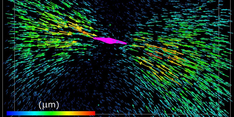
Wu and Hall’s colleagues used 3-D traction-force microscopy to measure the displacement of fluorescent marker beads distributed in a collagen matrix that also contained migrating breast-cancer cells. Shenoy’s team could then calculate the force exerted by the cells using the displacement of those beads.
“Nobody has looked how cellular forces quantitatively alter the matrix and how that feeds back into the cell force,” Shenoy said. “Using our model, we could quantitatively derive what forces the cells were exerting.”
The group published the findings in the Proceedings of the National Academy of Sciences.
Wu said the group’s work centered on a basic question: How much force do cells exert on their extracellular matrix when they migrate?
“The matrix is like a rope, and, in order for the cell to move, it has to exert force on this rope,” she said. “The question arose from cancer metastasis because, if the cells don’t move around, it’s a benign tumor and generally not life-threatening.”
It’s when the cancerous cell migrates that serious problems can arise. That migration occurs through “cross-talk” between the cell and the matrix, the group found. As the cell pulls on the matrix, the fibrous matrix stiffens; in turn, the stiffening of the matrix causes the cell to pull harder, which stiffens the matrix even more.
This increased stiffening also increases cell-force transmission distance, which can potentially promote metastasis of cancer cells.
“We’ve shown that the cells are able to align the fibers in their vicinity by exerting force,” Hall said. “We’ve also shown that, when the matrix is more fibrous, less like a continuous material and more like a mesh of fibers, they’re able to align the fibers through the production of force. And, once the fiber is aligned and taut, it’s easier for cells to pull on them and migrate.”
The combination of computer modeling and physical experiments helped to resolve confounding results in previous attempts to quantify cancerous cells’ rate of migration.
“This was a totally novel approach,” Shenoy said, “since it’s the first to account for the fact that collagen is fibrous. Without that model, you can’t explain the experimental data.”
This research was supported by grants from the National Institutes of Health, National Cancer Institute and National Science Foundation and made use of the Cornell NanoScale Science and Technology Facility, Cornell Biotechnology Resource Center Imaging Facility, Cornell Center for Materials Research and Cornell Nanobiotechnology Center.
Shenoy lab members Farid Alisafaei and Ehsan Ban also contributed to the study. Recently established through a Nation Science Foundation grant, Penn’s Center for Engineering Mechanobiology also supported this study.
